

Overview
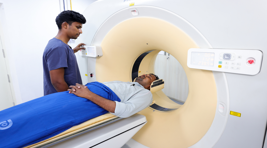
Magnetic Resonance Angiography (MRA) is a safe and non-invasive imaging technique that uses powerful magnetic fields, radio waves, and advanced computer technology to produce highly detailed images of blood vessels throughout the body. Unlike conventional angiography, MRA typically does not require catheter insertion, making it a more comfortable and lower-risk option for evaluating the vascular system.
MRA is commonly used to examine the arteries and veins in the brain, neck, heart, kidneys, abdomen, and limbs. It helps doctors detect and assess conditions such as narrowed or blocked arteries, aneurysms, blood clots, vascular malformations, and other circulatory disorders with exceptional accuracy. The detailed images provided by MRA enable early diagnosis, support personalized treatment planning, guide surgical or interventional procedures, and help monitor the effectiveness of ongoing treatment. Its ability to deliver precise vascular imaging without exposure to ionizing radiation makes MRA an important diagnostic tool in modern healthcare.
Technology Details
3D Vascular Imaging
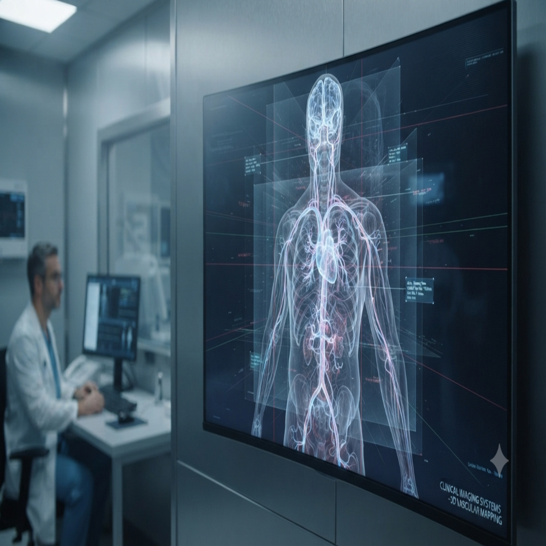
- Advanced MRA imaging creates highly detailed three-dimensional views of arteries and veins, allowing doctors to examine the vascular system with exceptional clarity.
- The enhanced visualization helps identify abnormalities such as narrowed or blocked blood vessels, aneurysms, blood clots, vascular malformations, and other circulatory disorders with greater accuracy.
- These precise images support early diagnosis, assist in treatment planning, guide interventional procedures when needed, and help monitor the effectiveness of ongoing vascular care.
Contrast-Enhanced MRA
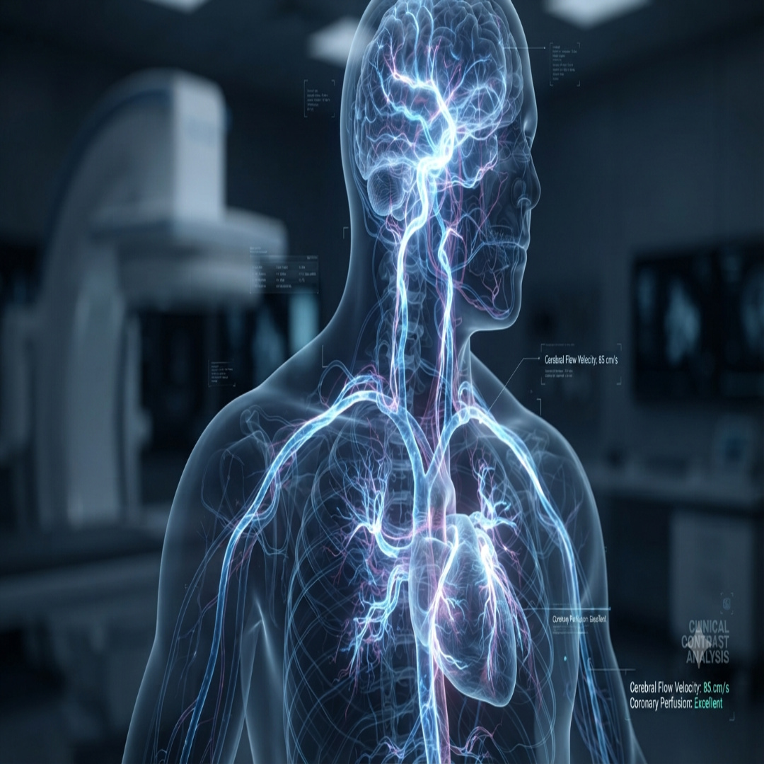
- When required, MRA scans use specialized contrast agents to enhance the visibility of blood vessels and improve the assessment of blood flow throughout the body.
- The contrast material helps produce clearer, more detailed images of arteries and veins, enabling doctors to accurately detect blockages, narrowing, aneurysms, vascular malformations, and other circulatory abnormalities.
- This enhanced imaging supports precise diagnosis, personalized treatment planning, and effective monitoring of vascular diseases while maintaining a minimally invasive approach.
When is this Technology Recommended?
This procedure is recommended for patients requiring advanced clinical interventions. Our specialists provide comprehensive assessments to determine eligibility.
Our health advisors will reach out to you
Request a Callback